Roustit M Blais Systematic Review of Secondary Raynaud
Short Review and Introduction
Raynaud's miracle (RP), first described in 1862 by Maurice Raynaud (Fardoun et al., 2016; Wigley and Flavahan, 2016), is nowadays in five–10% of the earth's population. It is a clinical consequence of recurrent vasospasm of the small-scale arteries and arterioles of the fingers and toes triggered by cold or even emotional stress (Wigley and Flavahan, 2016), at times also affecting the nose, ears, or lips (Block and Sequeira, 2001; Herrick, 2005; McMahan and Wigley, 2010; Hughes and Herrick, 2016). The skin usually turns white (ischemia), blue (deoxygenation) and then ruby (reperfusion) (Cake and Sequeira, 2001; Herrick, 2005; McMahan and Wigley, 2010; Hughes and Herrick, 2016).
There are 2 main categories, i.e., Primary (PRP) and Secondary RP (SRP) and most are PRP that have an isolated finding if there is no underlying pathology (idiopathic). SRP is present in various conditions, like connective tissue diseases (CTD), such as systemic sclerosis (SSc).
Ninety percent of SSc patients have RP which is the most common presenting feature and may precede diagnosis by many years (Cake and Sequeira, 2001; Herrick, 2005; McMahan and Wigley, 2010; Hughes and Herrick, 2016). The suggested criteria for PRP include symmetric attacks, the absence of tissue necrosis, ulceration or gangrene, the absence of a secondary cause, negative tests for antinuclear antibodies and a normal erythrocyte sedimentation rate (LeRoy and Medsger, 1992, 2001).
When PRP diagnosis is made no underlying disease has yet been identified, making prediction of if and when it may turn into SRP hard (Ingegnoli et al., 2010; Avouac et al., 2011; Bernero et al., 2013; Cutolo et al., 2017a). Nailfold video-capillaroscopy (NVC) is able to distinguish SRP from both PRP and healthy subjects past detecting morphological microcirculation abnormalities (Cake and Sequeira, 2001; Cutolo et al., 2003; Herrick, 2005; Ingegnoli et al., 2017; Pizzorni et al., 2017b; Herrick and Murray, 2018). Follow-upward nailfold capillaroscopic assay should be performed every 6 months in PRP patients (Cutolo et al., 2003; Bernero et al., 2013).
Nailfold capillaries in PRP are ordinarily normal in shape without any specific alterations (Ingegnoli et al., 2013; Smith et al., 2016a) or abnormal capillaroscopic findings, i.e., giant capillaries and microhemorrhages, whilst their presence is diagnostic for the "Early" NVC pattern of scleroderma microangiography (Cutolo et al., 2003, 2017a; Bernero et al., 2013; Ingegnoli et al., 2013; Smith et al., 2016a).
Indeed, abnormal nailfold capillaroscopic images (more specifically "scleroderma patterns") were included in the 2013 European League Against Rheumatism and American College of Rheumatology nomenclature criteria for SSc to this aim (van den Hoogen et al., 2013).
Digital vasculopathy is structural and functional in SRP due to SSc. NVC cannot measure blood perfusion (BP) nether standard conditions (Mugii et al., 2009) but other techniques, similar laser and thermography as well equally emerging technologies are able to evaluate and quantify skin blood flow and perfusion in SSc (Wigley et al., 1990; Clark et al., 2003; Murray et al., 2009; Rosato et al., 2009, 2011; Cutolo et al., 2010, 2014, 2018b; Pauling et al., 2012a,b, 2015; Della Rossa et al., 2013; Ruaro et al., 2014, 2016, 2017b, 2018c; Sulli et al., 2014; Lambrecht et al., 2016; Wilkinson et al., 2018). Laser Doppler flowmetry (LDF) evaluates blood menstruation at a single skin point, providing an alphabetize of skin perfusion (Cutolo et al., 2010, 2014; Ruaro et al., 2017b, 2018c).
Laser Doppler imaging (LDI) may likewise be used to evaluate the microcirculatory blood flow (Wigley et al., 1990; Clark et al., 2003; Murray et al., 2009; Rosato et al., 2009, 2011). LDI assesses more than ane area and is more effective than a unmarried probe Doppler (Wigley et al., 1990; Clark et al., 2003; Murray et al., 2009; Rosato et al., 2009, 2011). LDI can help to differentiate between PRP and patients with SRP to scleroderma (Wigley et al., 1990; Clark et al., 2003; Murray et al., 2009; Rosato et al., 2009, 2011). Although Murray et al. suggested that combining laser Doppler with other imaging modalities (e.g., nailfold capillaroscopy and thermal imaging) is more effective than laser Doppler solitary, these functional imaging tools are non however widely available (Murray et al., 2009).
Laser speckle contrast assay (LASCA) can quantify the blood flow over a divers area and is based on the principle that when laser light illuminates a tissue it forms a speckle pattern (Della Rossa et al., 2013; Ruaro et al., 2014; Lambrecht et al., 2016; Cutolo et al., 2018b). Changes in this pattern are analyzed past software and the static areas show a stationary speckle design, in contrast with the moving objects like reddish claret cells that crusade the speckle pattern to fluctuate and appear blurred. The level of blurring (contrast) is analyzed and interpreted every bit BP (Cutolo et al., 2017a; Ingegnoli et al., 2017). LASCA is a fast imaging technique, with a high resolution and reliability, as recently demonstrated in two studies (Lambrecht et al., 2016; Cutolo et al., 2018b).
LASCA has been applied in research studies on RP and SSc (Della Rossa et al., 2013; Ingegnoli et al., 2013, 2017) and one demonstrated that peripheral BP evaluated by both LDF and LASCA correlates to the extent of the microangiopathy (Ruaro et al., 2014).
Laser speckle dissimilarity imaging (LCSI) is similar to LASCA and provides a five-fold increase in spatial resolution over LASCA. However, it is more time consuming (Pauling et al., 2015).
Thermal imaging (TI), an indirect method, makes apply of a thermal camera to image the pare temperature to show the underlying blood flow (Clark et al., 2003; Murray et al., 2009; Pauling et al., 2012a,b; Wilkinson et al., 2018). TI evaluated RP in several studies and the response to lower temperatures (common cold) was able to differentiate betwixt PRP and SRP to SSc (Murray et al., 2009). However, it has a poor sensitivity in detecting BP variations and has a low spatial resolution (Murray et al., 2009).
Non-invasive assessment of the morphological and functional peripheral apportionment may supplement the concrete examination and provide a quick, accurately diagnosis, ultimately guiding the correct treatment for both PRP and SRP (Filaci et al., 1999, 2001; Faggioli et al., 2006; Pyrpasopoulou and Aslanidis, 2007; Aschwanden et al., 2008; Caramaschi et al., 2009; Miniati et al., 2009; Shah et al., 2011; Guiducci et al., 2012; Roustit et al., 2012; Cutolo et al., 2013, 2017b; Herrick, 2013, 2017; Cutolo and Sulli, 2015; Gladue et al., 2016; Smith et al., 2016b; Trombetta et al., 2016; Burmester et al., 2017; Kowal-Bielecka et al., 2017; Ruaro et al., 2017a; Rotondo et al., 2018).
Most PRP patients accept no serious symptoms and respond well to conservative non-medical treatment like keeping warm and fugitive drugs with vasoconstrictive effects. Whilst other cases require pharmacological handling similar calcium aqueduct blockers as first-line therapy (Herrick, 2013). Although diverse treatment options are available for the management of SSc-related SRP, these approaches at nearly reduce the severity of the symptoms just do not resolve the clinical state of affairs (Herrick, 2013; Cutolo and Sulli, 2015; Gladue et al., 2016; Herrick, 2017; Kowal-Bielecka et al., 2017).
The revised European League Against Rheumatism (EULAR) recommendations for RP in SSc patients (SSc-RP) handling state that "calcium aqueduct blockers should exist used every bit first-line therapy and PDE-5 inhibitors in patients with SSc with astringent RP and/or those who do not satisfactorily answer to calcium channel blockers" (Kowal-Bielecka et al., 2017). The experts recommended that "intravenous prostanoids are considered when oral therapies (including calcium channel blockers and PDE-5 inhibitors) have failed" and they likewise recognize that "fluoxetine is a useful alternative for treatment of SSc-RP, in particular in patients with SSc who cannot tolerate or do not respond to vasodilators" (Kowal-Bielecka et al., 2017).
As aforementioned, the current therapies for RP are often ineffective. Therefore, the biggest challenge is identifying a drug able to halt RP progression or ameliorate yet, to preclude the microvascular anomalies which involve tissue hypoperfusion and hypoxia.
That is why an NVC-based assessment of microvascular structure and an evaluation of functional impairment by laser tools and thermography may be useful to assess the efficacy of pharmacological therapies during the treatment of RP patients.
Interestingly, some studies used NVC to detect the microvascular changes as possible markers of response to immunosuppressive/anti-fibrosing handling and vasoactive drugs (Filaci et al., 1999, 2001; Faggioli et al., 2006; Pyrpasopoulou and Aslanidis, 2007; Caramaschi et al., 2009; Miniati et al., 2009; Shah et al., 2011; Guiducci et al., 2012; Cutolo et al., 2013; Smith et al., 2016b; Trombetta et al., 2016; Ruaro et al., 2017a). Early studies on the upshot of Cyclosporin have shown a moderate comeback in clinical symptoms and SSc nailfold microangiopathy, after a 12 month treatment cycle (Filaci et al., 2001; Caramaschi et al., 2009).
Similarly, Cyclophosphamide administration was reported to be significantly associated with an comeback in microvascular damage and a regression of the capillaroscopic pattern severity (Caramaschi et al., 2009).
A recent report showed no progression (therefore a positive affliction modifying effect) of the microvascular damage (mainly no farther capillary loss) during the 12-calendar month follow-up in patients with early SSc and diffuse skin interest treated with Rituximab (Smith et al., 2016b).
Contempo studies have reported that the use of autologous haemopoietic stem cell transplantation in patients with severe diffuse SSc improved microangiopathy and the NVC blueprint changed from "Tardily" to "Agile" (Miniati et al., 2009). Three studies reported an comeback in nailfold microvascularization afterwards iloprost handling (Faggioli et al., 2006; Pyrpasopoulou and Aslanidis, 2007; Shah et al., 2011; Rotondo et al., 2018). Various studies used NVC with laser techniques to access the drug response in SSc patients treated with a combination of intravenous prostanoids and endothelin-1 receptor blockers, reporting a pregnant capillary loss reduction (Guiducci et al., 2012; Cutolo et al., 2013, 2014, 2016; Trombetta et al., 2016; Ruaro et al., 2017a).
The objectives of this study were:
(i) to provide a brusque review in the introduction on morphological (NVC) and functional techniques (laser tools and thermography) that allow for a right early diagnosis and handling of main and PRP;
(ii) to nowadays a pilot report that compares BP measured by LASCA in unlike skin areas of the easily and confront in patients with PRP, SRP to SSc and healthy subjects (CNT).
Patients and Methods of the Airplane pilot Study
Written report Population
A full of 31 PRP patients were enrolled after having obtained their written informed consent for the employ of imaging and the demographic data as educational material and for publications.
All the PRP patients fulfilled the LeRoy criteria (LeRoy and Medsger, 2001) as did 68 SSc patients, who met the ACR/EULAR 2013 criteria for SSc (van den Hoogen et al., 2013) during routine clinical assessment in our Rheumatology Section, from October, 2016 to Mach, 2017. The study was carried out co-ordinate to the ethical standard of Skilful Clinical Do. A consummate medical history was collected and all participants had a clinical examination (Table 1).
Table i. Clinical findings in patients with primary Raynaud's phenomenon (PRP), systemic sclerosis (SSc) and healthy subjects (CNT).
The inclusion criteria were a diagnosis of PRP or SRP to SSc, and all patients had been on a stable drug regimen for at to the lowest degree 2 months prior study entry.
The exclusion criterion was existence on a drug regimen that could potentially influence blood menstruation.
If the patients were existence treated with prostanoids and endothelin-1 receptor antagonists, they were temporarily withdrawn 1 month earlier instrumental assessment.
All SSc patients were taking aspirin (average dosage 100 mg/solar day) at the fourth dimension of the study. Other concomitant treatment included: proton pump inhibitors (used by #52 patients), antihypertensive drugs i.eastward., angiotensin-converting enzyme (ACE) inhibitors (#9 patients), cyclosporine (average dosage 150 mg/day: #12 patients), methotrexate (average dosage seven.5 mg/calendar week: #12 patients). The PRP therapy treatment was: proton pump inhibitors (used by #8 patients), antihypertensive drugs i.eastward., ACE inhibitors (#three patients).
Both LASCA and NVC were performed on the same 24-hour interval in all PRP and SSc patients.
Light amplification by stimulated emission of radiation speckle contrast assay was also performed in the 70 healthy subjects (CNT) matched with the RP patients for age and gender (run into Tabular array 1 for demographic data).
Laser Speckle Contrast Analysis (LASCA)
Skin BP was analyzed by the LASCA technique (Pericam PSI, Perimed, Milan, Italy) at the level of dorsal and palmar attribute of easily and the whole face, in both SSc patients and healthy subjects every bit previously described (Ruaro et al., 2014, 2016; Sulli et al., 2014). Dissimilar regions of involvement (ROIs) were created, as previously reported, i.eastward., at the level of fingertips, periungual areas, dorsal and palmar aspect of the tertiary finger bilaterally, the dorsum and palm of both hands and confront (forehead, tip of nose, zygomas and perioral region) (see Figure 1 for ROI areas) (Sulli et al., 2014; Ruaro et al., 2016, 2018b).
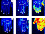
Figure i. Laser Speckle Contrast Analysis (LASCA) images of secondary Raynaud's phenomenon (RP) to systemic sclerosis, in a patient with a "Late" pattern of scleroderma microangiopathy (A), primary RP (B) and a healthy subject (C), showing the regions of interest (ROI - white circles) created at the level of back and palm of the hand, dorsal and palmar aspect of the 3rd finger, periungual areas and fingertips to evaluate blood perfusion. Color code: blue corresponds to a low BP, xanthous an intermediate BP and red a higher BP. Noteworthy is the fact that subjects with a late design have a prevalence of blue, indicating a low perfusion level.
The average BP from either fingertips or periungual areas was calculated past summing the perfusion values of eight fingers together then dividing the final value by the number of fingers.
The average BP from the two palmar and dorsal areas of the fingers, palm and dorsum of the hands and zygoma was calculated by summing the perfusion values of the 2 sides (correct and left) and so dividing the last value by two. The BP was quantified as perfusion units (PU; Sulli et al., 2014; Ruaro et al., 2016). The aforementioned operator (BR) performed the examination in all PRP, SRP-SSc patients and CNT.
Nailfold Videocapillaroscopy (NVC)
All patients were assessed by nailfold videocapillaroscopy (NVC), (equipped with a 200× contact lens, connected to image assay software – Videocap, DS MediGroup, Milan, Italy) so as to distinguish PRP from SRP and to make up one's mind the correct nailfold microangiopathy pattern ("Early," "Agile," or "Late" pattern, co-ordinate to the Cutolo's criteria) in the SSc patients (Sulli et al., 2008; Smith et al., 2010, 2013; Table i). The aforementioned operator (CP) performed the examination in all PRP and SRP-SSc patients and CNT.
Statistical Analysis
The statistical analysis was carried out by parametric procedures and confirmed by non-parametric tests. The Mann-Whitney U test was performed to compare unpaired groups of variables, along with the Kruskal-Wallis test to compare continuous variables with nominal variables that had more than two levels. Any p-values beneath 0.05 were considered statistically pregnant. The results are given every bit, median and interquartile range (IQR).
Results
Both PRP and SSc patients had statistically significant lower BP than the healthy subjects at the fingertip (p < 0.0001), the periungual area (p < 0.0001), the palmar aspect of the third finger (p < 0.0001) and the palm areas (p < 0.0001). Conversely, all three groups had similar BP values in the other areas of the manus (dorsal aspect of the 3rd finger and dorsum of hand) and face (forehead, tip of nose, zygomas and perioral region). Moreover, BP was statistically significantly lower in PRP than in SSc patients with the "Early" blueprint of microangiopathy at fingertip (p = 0.04), periungual (p < 0.05), palmar attribute of the 3rd finger (p = 0.0008) and the palm areas (p = 0.0009). No statistically pregnant difference was observed between PRP and the "Early" design of microangiopathy in the other areas evaluated.
A statistically significant progressive decrease in BP was confirmed in SSc patients with a progressive pattern of nailfold microangiopathy ("Early," "Active," and "Belatedly") at the fingertip, periungual, palmar aspect of the third fingers and palm areas (p < 0.05). No statistically pregnant difference was observed between NVC patterns and BP at the level of the other areas (back of easily, whole face and different areas of face up) (p > 0.05) (Tabular array 2).
Table ii. Blood perfusion (BP) in systemic sclerosis (SSc), principal Raynaud's phenomenon (PRP) and healthy subjects (CNT).
If the iii nailfold microangiopathy patterns ("Early," "Active," and "Late") are evaluated separately, there is a statistically pregnant difference only between the "Early" and "Late" group, at the level of the fingertip, periungual, palmar aspect of the 3rd fingers and palm areas (p < 0.05). No statistically significant difference was observed in the other areas.
At that place were very few smokers in our written report and there was no statistically pregnant difference in the smoking habit between the groups.
Word
Our pilot study shows that the hand BP, evaluated by LASCA, was lower in PRP than in SSc patients with an "Early" NVC microangiopathy pattern.
The results of this report also confirm that SSc patients had a significant lower median BP than healthy subjects and the progressive subtract of BP in SSc patients with unlike: "Early," "Active," or "Late" NVC pattern of microangiopathy at the level of hand.
Indeed, some authors have reported different perfusion values in PRP and SRP to SSc patients, but the perfusion was evaluated either after, or during, different forms of stress, such every bit the cold or occlusion test, in contrast with our study where the perfusion was evaluated at basal condition (Pauling et al., 2012a, 2015).
Nosotros would like to attest that all the PRP patients had a functional disorder/dysfunction in microvascular circulation and our data emphasize the importance of the perfusion reduction, even in a functional phenomenon such as in PRP patients.
Moreover, our data are in understanding with those of other studies that report NVC every bit being the all-time method to evaluate microcirculation morphological and permanent damage and to make a differential diagnosis betwixt PRP and SRP (Murray et al., 2009; Ingegnoli et al., 2017; Herrick and Murray, 2018).
As previously reported our information confirm that patients with the "Late" SSc microangiopathy blueprint had a lower blood flow than those with the "Active" or "Early" SSc patterns at NVC (Ruaro et al., 2014, 2018b). In our precedent article we too reported that when BP was assessed by the LASCA technique significantly lower values were observed in the SSc patients than in the healthy subjects at the level of the fingertips, periungual areas and palm of the hands, with a statistically significant negative correlation between the extent of the nailfold microangiopathy and the BP values at the level of the same skin areas in SSc patients (Ruaro et al., 2014, 2018b).
The increased interest in microcirculation has led to a rapid development of new assessment methods. However, these techniques lack the support of validation studies as to their application in clinical practice. Nevertheless, microvascular structure evaluation by NVC combined with functional investigation by laser techniques or TI, non only helps in the distinction between primary and SRP, simply is also able to evaluate therapy response and illness progression (Filaci et al., 2001; Caramaschi et al., 2009; Guiducci et al., 2012; Cutolo et al., 2013, 2014, 2016; Smith et al., 2013, 2016b; Trombetta et al., 2016; Ruaro et al., 2017a, 2018a, Pizzorni et al., 2017a; Soulaidopoulos et al., 2017; Markusse et al., 2017).
In particular, the assessment of the number of capillary changes seems the all-time validated NVC parameter and is today evaluable with automated systems (Cutolo et al., 2018a).
In summary we are of the stance that morphological evaluation by NVC is the all-time method for the early on detection and quantification of microvascular abnormalities that characterize SRP. We also believe that clinicians should non underestimate RP which should have a scheduled follow-up as it might well exist a precocious cloaked clinical sign of abnormal microcirculation and a risk factor for the development of a CTD, especially SSc.
Last but not least, the primary message of this work is that while today there is no curative treatment all RP patients, because it is a very heterogeneous miracle, still there are many treatment options to amend quality of life of these patients. The early on detection of disease and immediate intervention appears to make a difference, such likewise-designed clinical trials and collaboration with networks, such as the European Reference Network on Rare and Circuitous Connective Tissue and Musculoskeletal Diseases Project and specialized centers carrying the research in this field with the aim of defining ideal diagnostic and therapeutic options (Smith et al., 2018).
Ethics Argument
This study has been performed in accord with the ethical standards laid downwards in the 1964 Proclamation of Helsinki and its later on amendments. Ethics approval was obtained from the local Upstanding Board and all patients gave written informed consent to enter the study.
Author Contributions
All authors listed have fabricated a substantial, direct and intellectual contribution to the work, and approved it for publication.
Funding
This written report was supported by funding from the Research Laboratory and Academic Division of Clinical Rheumatology of the University of Genova, Italia.
Conflict of Interest Statement
The authors declare that the research was conducted in the absence of any commercial or financial relationships that could be construed as a potential conflict of interest.
Acknowledgments
The authors would like to give thanks Barbara Wade, contract Professor at the University of Turin, for her linguistic advice. BR was supported by an EULAR scientific training bursary. VS is a Senior Clinical Investigator of the Inquiry Foundation – Flemish region (Belgium) (FWO) (1802915N).
References
Aschwanden, M., Daikeler, T., Jaeger, K. A., Thalhammer, C., Gratwohl, A., Matucci-Cerinic, M., et al. (2008). Rapid improvement of nailfold capillaroscopy later intense immunosuppression for systemic sclerosis and mixed tissue affliction. Ann. Rheum. Dis. 67, 1057–1059. doi: 10.1136/ard.2007.082008
PubMed Abstract | CrossRef Total Text | Google Scholar
Avouac, J., Fransen, J., Walker, U. A., Riccieri, V., Smith, 5., Muller, C., et al. (2011). Preliminary criteria for the very early diagnosis of systemic sclerosis: results of a delphi consensus study from EULAR scleroderma trials and research group. Ann. Rheum. Dis. 70, 476–481. doi: 10.1136/ard.2010.136929
PubMed Abstract | CrossRef Full Text | Google Scholar
Bernero, E., Sulli, A., Ferrari, G., Ravera, F., Pizzorni, C., Ruaro, B., et al. (2013). Prospective capillaroscopy-based study on transition from primary to secondary Raynaud's phenomenon: preliminary results. Reumatismo 65, 186–191. doi: x.4081/reumatismo.2013.186
PubMed Abstract | CrossRef Full Text | Google Scholar
Burmester, G. R., Bijlsma, J. Westward. J., Cutolo, M., and McInnes, I. B. (2017). Managing rheumatic and musculoskeletal diseases - past, present and future. Nat. Rev. Rheumatol. 13, 443–448. doi: ten.1038/nrrheum.2017.95
PubMed Abstruse | CrossRef Total Text | Google Scholar
Caramaschi, P., Volpe, A., Pieropan, South., Tinazzi, I., Mahamid, H., Bambara, L. Grand., et al. (2009). Cyclophosphamide handling improves microvessel damage in systemic sclerosis. Clin. Rheumatol. 28, 391–395. doi: ten.1007/s10067-008-1058-y
PubMed Abstract | CrossRef Full Text | Google Scholar
Clark, S., Dunn, G., Moore, T., Jayson, M., King, T. A., and Herrick, A. 50. (2003). Comparing of thermography and light amplification by stimulated emission of radiation Doppler imaging in the assessment of Raynaud'due south phenomenon. Microvasc. Res. 66, 73–76. doi: ten.1016/S0026-2862(03)00018-9
CrossRef Total Text | Google Scholar
Cutolo, M., Damjanov, N., Ruaro, B., Zekovic, A., and Smith, V. (2016). Imaging of connective tissue diseases: beyond visceral organ imaging? All-time Pract. Res. Clin. Rheumatol. 30, 670–687. doi: 10.1016/j.berh.2016.10.002
PubMed Abstract | CrossRef Full Text | Google Scholar
Cutolo, M., Ferrone, C., Pizzorni, C., Soldano, S., Seriolo, B., and Sulli, A. (2010). Peripheral blood perfusion correlates with microvascular abnormalities in systemic sclerosis: a laser-Doppler and nailfold videocapillaroscopy report. J. Rheumatol. 37, 1174–1180. doi: x.3899/jrheum.091356
PubMed Abstruse | CrossRef Full Text | Google Scholar
Cutolo, Chiliad., Ruaro, B., Pizzorni, C., Ravera, F., Smith, V., Zampogna, G., et al. (2014). Longterm handling with endothelin receptor antagonist bosentan and iloprost improves fingertip claret perfusion in systemic sclerosis. J. Rheumatol. 41, 881–886. doi: 10.3899/jrheum.131284
PubMed Abstract | CrossRef Full Text | Google Scholar
Cutolo, K., Smith, V., Distler, O., Kowal-Bielecka, O., Allanore, Y., and Matucci-Cerinic, G. (2017a). Preliminary analysis of nailfold capillaroscopy in very early diagnosis of systemic sclerosis (VEDOSS): the CAPI-VEDOSS experience. Ann. Rheum. Dis. 76, 65–66.
Google Scholar
Cutolo, M., Smith, 5., Furst, D. East., Khanna, D., and Herrick, A. L. (2017b). Points to consider-Raynaud's miracle in systemic sclerosis. Rheumatology 56, 45–48. doi: 10.1093/rheumatology/kex199
PubMed Abstract | CrossRef Total Text | Google Scholar
Cutolo, M., and Sulli, A. (2015). Therapy: optimized handling algorithms for digital vasculopathy in systemic sclerosis. Nat. Rev. Rheumatol. 11, 569–571. doi: ten.1038/nrrheum.2015.111
PubMed Abstract | CrossRef Full Text | Google Scholar
Cutolo, One thousand., Trombetta, A. C., Melsens, K., Pizzorni, C., Sulli, A., Ruaro, B., et al. (2018a). Automated assessment of absolute nailfold capillary number on videocapillaroscopic images: proof of principle and validation in systemic sclerosis. Microcirculation 25:e12447. doi: 10.1111/micc.12447
PubMed Abstract | CrossRef Total Text | Google Scholar
Cutolo, G., Vanhaecke, A., Ruaro, B., Deschepper, E., Ickinger, C., Melsens, G., et al. (2018b). EULAR study group on microcirculation in rheumatic diseases. Is laser speckle contrast analysis (LASCA) the new kid on the block in systemic sclerosis? A systematic literature review and airplane pilot study to evaluate reliability of LASCA to mensurate peripheral blood perfusion in scleroderma patients. Autoimmun. Rev. 17, 775–780. doi: ten.1016/j.autrev.2018.01.023
PubMed Abstract | CrossRef Full Text | Google Scholar
Cutolo, M., Zampogna, G., Vremis, L., Smith, Five., Pizzorni, C., and Sulli, A. (2013). Longterm effects of endothelin receptor antagonism on microvascular damage evaluated past nailfold capillaroscopic analysis in systemic sclerosis. J. Rheumatol. forty, forty–45. doi: ten.3899/jrheum.120416
PubMed Abstract | CrossRef Total Text | Google Scholar
Della Rossa, A., Cazzato, M., d'Ascanio, A., Tavoni, A., Bencivelli, Westward., Pepe, P., et al. (2013). Amending of microcirculation is a authentication of very early systemic sclerosis patients: a laser speckle dissimilarity assay. Clin. Exp. Rheumatol. 31, S109–S114.
PubMed Abstract | Google Scholar
Faggioli, P., Giani, Fifty., and Mazzone, A. (2006). Possible role of iloprost (stable analogue of PGI2) in promoting neoangiogenesis in systemic sclerosis. Clin. Exp. Rheumatol. 24, 220–221.
Google Scholar
Fardoun, M. M., Nassif, J., Issa, Thousand., Baydoun, Due east., and Eid, A. H. (2016). Raynaud's phenomenon: a brief review of the underlying mechanisms. Front. Pharmacol. 7:438. doi: 10.3389/fphar.2016.00438
CrossRef Total Text | Google Scholar
Filaci, G., Cutolo, Thousand., Basso, 1000., Murdaca, M., Derchi, 50., Gianrossi, R., et al. (2001). Long-term handling of patients affected past systemic sclerosis with cyclosporin A. Rheumatology 40, 1431–1434. doi: 10.1093/rheumatology/40.12.1431
CrossRef Total Text | Google Scholar
Filaci, G., Cutolo, Yard., Scudeletti, M., Castagneto, C., Derchi, 50., Gianrossi, R., et al. (1999). Cyclosporin A and iloprost treatment of systemic sclerosis: clinical results and interleukin-6 serum changes afterwards 12 months of therapy. Rheumatology 38, 992–996. doi: 10.1093/rheumatology/38.10.992
PubMed Abstract | CrossRef Full Text | Google Scholar
Gladue, H., Berrocal, V., Harris, R., Tsou, P. S., Edhayan, G., Ohara, R., et al. (2016). A randomized controlled trial of acupressure for the treatment of Raynaud's miracle: the difficulty of conducting a trial in Raynaud's phenomenon. J. Scleroderma Relat. Disord. i, 226–233. doi: 10.5301/jsrd.5000206
PubMed Abstract | CrossRef Full Text | Google Scholar
Guiducci, S., Bellando Randone, South., Bruni, C., Carnesecchi, G., Maresta, A., Iannone, F., et al. (2012). Bosentan fosters microvascular de-remodelling in systemic sclerosis. Clin. Rheumatol. 31, 1723–1725. doi: 10.1007/s10067-012-2074-5
PubMed Abstruse | CrossRef Full Text | Google Scholar
Herrick, A. L., and Murray, A. (2018). The office of capillaroscopy and thermography in the cess and management of Raynaud's phenomenon. Autoimmun. Rev. 17, 465–472. doi: ten.1016/j.autrev.2017.11.036
PubMed Abstract | CrossRef Full Text | Google Scholar
Ingegnoli, F., Boracchi, P., Gualtierotti, R., Biganzoli, E. M., Zeni, S., Lubatti, C., et al. (2010). Improving outcome prediction of systemic sclerosis from isolated Raynaud's phenomenon: role of autoantibodies and nail-fold capillaroscopy. Rheumatology 49, 797–805. doi: 10.1093/rheumatology/ kep447
PubMed Abstruse | CrossRef Full Text | Google Scholar
Ingegnoli, F., Gualtierotti, R., Lubatti, C., Bertolazzi, C., Gutierrez, Thou., Boracchi, P., et al. (2013). Nailfold capillary patterns in salubrious subjects: a real event in capillaroscopy. Microvasc. Res. xc, ninety–95. doi: 10.1016/j.mvr.2013.07.001
PubMed Abstract | CrossRef Full Text | Google Scholar
Ingegnoli, F., Ughi, N., Dinsdale, Chiliad., Orenti, A., Boracchi, P., Allanore, Y., et al. (2017). An international SUrvey on non-iNvaSive tecHniques to assess the mIcrocirculation in patients with RayNaud's miracle (SUNSHINE survey). Rheumatol. Int. 37, 1879–1890. doi: 10.1007/s00296-017-3808-0
PubMed Abstract | CrossRef Total Text | Google Scholar
Kowal-Bielecka, O., Fransen, J., Avouac, J., Becker, M., Kulak, A., Allanore, Y., et al. (2017). Update of EULAR recommendations for the treatment of systemic sclerosis. Ann. Rheum. Dis. 76, 1327–1339. doi: x.1136/annrheumdis-2016-209909
PubMed Abstract | CrossRef Full Text | Google Scholar
Lambrecht, V., Cutolo, M., De Keyser, F., Decuman, South., Ruaro, B., Sulli, A., et al. (2016). Reliability of the quantitative assessment of peripheral claret perfusion by light amplification by stimulated emission of radiation speckle contrast analysis in a systemic sclerosis cohort. Ann. Rheum. Dis. 75, 1263–1264. doi: ten.1136/annrheumdis-2015-208857
PubMed Abstract | CrossRef Total Text | Google Scholar
LeRoy, E. C., and Medsger, T. A. (1992). Raynaud's phenomenon: a proposal for classification. Clin. Exp. Rheumatol. 10, 485–488.
Google Scholar
LeRoy, E. C., and Medsger, T. A. (2001). Criteria for the nomenclature of early systemic sclerosis. J. Rheumatol. 28, 1573–1576.
Google Scholar
Markusse, I. M., Meijs, J., de Boer, B., Bakker, J. A., Schippers, H. P. C., Schouffoer, A. A., et al. (2017). Predicting cardiopulmonary involvement in patients with systemic sclerosis: complementary value of nailfold videocapillaroscopy patterns and disease-specific autoantibodies. Rheumatology 56, 1081–1088. doi: ten.1093/rheumatology/kew402
PubMed Abstract | CrossRef Full Text | Google Scholar
McMahan, Z. H., and Wigley, F. M. (2010). Raynaud's phenomenon and digital ischemia: a practical approach to risk stratification, diagnosis and management. Int. J. Clin. Rheumtol. 5, 355–370. doi: 10.2217/ijr.10.17
CrossRef Total Text | Google Scholar
Miniati, I., Guiducci, S., Conforti, Grand. L., Rogai, V., Fiori, M., Cinelli, M., et al. (2009). Autologous stem jail cell transplantation improves microcirculation in systemic sclerosis. Ann. Rheum. Dis. 68, 94–98. doi: 10.1136/ard.2007.082495
PubMed Abstract | CrossRef Full Text | Google Scholar
Mugii, N., Hasegawa, M., Hamaguchi, Y., Tanaka, C., Kaji, M., Komura, M., et al. (2009). Reduced red blood cell velocity in nailfold capillaries as a sensitive and specific indicator of microcirculation injury in systemic sclerosis. Rheumatology 48, 696–703. doi: ten.1093/rheumatology/kep066
PubMed Abstruse | CrossRef Full Text | Google Scholar
Murray, A. K., Moore, T. L., Manning, J. B., Taylor, C., Griffiths, C. E., and Herrick, A. L. (2009). Noninvasive imaging techniques in the assessment of scleroderma spectrum disorders. Arthr. Rheum. 61, 1103–1111. doi: 10.1002/art.24645
PubMed Abstruse | CrossRef Full Text | Google Scholar
Pauling, J. D., Shipley, J. A., Harris, N. D., and McHugh, N. J. (2012a). Use of infrared thermography equally an endpoint in therapeutic trials of Raynaud's phenomenon and systemic sclerosis. Clin. Exp. Rheumatol. 30, S103–S115.
PubMed Abstruse | Google Scholar
Pauling, J. D., Shipley, J. A., Raper, S., Watson, Thousand. L., Ward, S. G., Harris, N. D., et al. (2012b). Comparison of infrared thermography and laser speckle dissimilarity imaging for the dynamic cess of digital microvascular office. Microvasc. Res. 83, 162–167. doi: ten.1016/j.mvr.2011.06.012
PubMed Abstract | CrossRef Total Text | Google Scholar
Pauling, J. D., Shipley, J. A., Hart, D. J., McGrogan, A., and McHugh, N. J. (2015). Apply of laser speckle dissimilarity imaging to assess digital microvascular part in primary raynaud phenomenon and systemic sclerosis: a comparing using the raynaud status score diary. J. Rheumatol. 42, 1163–1168. doi: 10.3899/jrheum.141437
PubMed Abstract | CrossRef Full Text | Google Scholar
Pizzorni, C., Sulli, A., Paolino, S., Ruaro, B., Smith, V., Trombetta, A. C., et al. (2017a). Progression of organ involvement in systemic sclerosis patients with persistent "Tardily" nailfold capillaroscopic design of microangiopathy: a prospective study. J. Rheumatol. 44, 1941–1942. doi: 10.3899/jrheum.170485
PubMed Abstract | CrossRef Full Text | Google Scholar
Pizzorni, C., Sulli, A., Smith, V., Ruaro, B., Trombetta, A. C., Cutolo, K., et al. (2017b). Primary Raynaud's phenomenon and nailfold videocapillaroscopy: age-related changes in capillary morphology. Clin. Rheumatol. 36, 1637–1642. doi: 10.1007/s10067-016-3442-iii
PubMed Abstruse | CrossRef Full Text | Google Scholar
Rosato, E., Borghese, F., Pisarri, S., and Salsano, F. (2009). Laser Doppler perfusion imaging is useful in the study of Raynaud's miracle and improves the capillaroscopic diagnosis. J. Rheumatol. 36, 2257–2263. doi: 10.3899/jrheum.090187
PubMed Abstract | CrossRef Full Text | Google Scholar
Rosato, E., Rossi, C., Molinaro, I., Giovannetti, A., Pisarri, S., and Salsano, F. (2011). Laser Doppler perfusion imaging in systemic sclerosis impaired response to common cold stimulation involves digits and hand dorsum. Rheumatology fifty, 1654–1658. doi: 10.1093/rheumatology/ker188
PubMed Abstract | CrossRef Full Text | Google Scholar
Rotondo, C., Nivuori, M., Chialà, A., Praino, E., Matucci Cerinic, M., Cutolo, M., et al. (2018). Evidence for increase in finger blood period, evaluated by laser Doppler flowmetry, following iloprost infusion in patients with systemic sclerosis: a week-long observational longitudinal report. Scand. J. Rheumatol. 47, 311–318. doi: 10.1080/03009742.2017.1397187
PubMed Abstract | CrossRef Full Text | Google Scholar
Roustit, One thousand., Hellmann, M., Cracowski, C., Blaise, Southward., and Cracowski, J. 50. (2012). Sildenafil increases digital peel blood flow during all phases of local cooling in main Raynaud's phenomenon. Clin. Pharmacol. Ther. 91, 813–819. doi: 10.1038/clpt.2011.302
PubMed Abstract | CrossRef Full Text | Google Scholar
Ruaro, B., Casabella, A., Paolino, South., Pizzorni, C., Alessandri, E., Seriolo, C., et al. (2018a). Correlation between bone quality and microvascular damage in systemic sclerosis patients. Rheumatology 57, 1548–1554. doi: x.1093/rheumatology/key130
PubMed Abstruse | CrossRef Total Text | Google Scholar
Ruaro, B., Sulli, A., Pizzorni, C., Paolino, S., Smith, V., Alessandri, E., et al. (2018b). Correlations between claret perfusion and dermal thickness in different pare areas of systemic sclerosis patients. Microvasc. Res. 115, 28–33. doi: 10.1016/j.mvr.2017.08.004
PubMed Abstract | CrossRef Total Text | Google Scholar
Ruaro, B., Sulli, A., Smith, V., Pizzorni, C., Paolino, S., Alessandri, E., et al. (2018c). Advances in nailfold capillaroscopic assay in systemic sclerosis. JSRD iii, 122–131. doi: 10.1136/bmjopen-2017-020479
PubMed Abstract | CrossRef Total Text | Google Scholar
Ruaro, B., Paolino, S., Pizzorni, C., Cutolo, M., and Sulli, A. (2017a). Cess of handling effects on digital ulcer and claret perfusion by laser speckle dissimilarity analysis in a patient afflicted by systemic sclerosis. Reumatismo 2017, 134–136. doi: x.4081/reumatismo.2017.986
PubMed Abstruse | CrossRef Full Text | Google Scholar
Ruaro, B., Sulli, A., Smith, 5., Pizzorni, C., Paolino, South., Alessandri, E., et al. (2017b). Microvascular damage evaluation in systemic sclerosis: the role of nailfold videocapillaroscopy and light amplification by stimulated emission of radiation techniques. Reumatismo. 69, 147–155. doi: x.4081/reumatismo.2017.959
PubMed Abstract | CrossRef Full Text | Google Scholar
Ruaro, B., Sulli, A., Pizzorni, C., Paolino, S., Smith, V., and Cutolo, Yard. (2016). Correlations between skin blood perfusion values and nailfold capillaroscopy scores in systemic sclerosis patients. Microvasc. Res. 105, 119–124. doi: 10.1016/j.mvr.2016.02.007
PubMed Abstruse | CrossRef Full Text | Google Scholar
Ruaro, B., Sulli, A., Smith, 5., Pizzorni, C., Gallo, M., and Cutolo, M. (2014). Light amplification by stimulated emission of radiation speckle contrast analysis: a new method to evaluate peripheral blood perfusion in systemic sclerosis patients. Ann. Rheum. Dis. 73, 1181–1185. doi: 10.1136/annrheumdis-2013-203514
PubMed Abstract | CrossRef Full Text | Google Scholar
Shah, P., Murray, A. 1000., Moore, T. Fifty., and Herrick, A. Fifty. (2011). Effects of iloprost on microvascular structure assessed past nailfold videocapillaroscopy: a pilot study. J. Rheumatol. 38, 2079–2080. doi: 10.3899/jrheum.110067
PubMed Abstruse | CrossRef Total Text | Google Scholar
Smith, V., Beeckman, S., Herrick, A. L., Decuman, S., Deschepper, E., De Keyser, F., et al. (2016a). An EULAR written report group pilot study on reliability of simple capillaroscopic definitions to describe capillary morphology in rheumatic diseases. Rheumatology 55, 883–890. doi: 10.1093/rheumatology/kev441
PubMed Abstract | CrossRef Full Text | Google Scholar
Smith, V., Pizzorni, C., Riccieri, V., Decuman, S., Brusselle, Thousand., De Pauw, M., et al. (2016b). Stabilization of microcirculation in patients with early systemic sclerosis with diffuse skin interest following rituximab treatment: an open up-characterization Study. J. Rheumatol. 43, 995–996. doi: 10.3899/jrheum.151018
PubMed Abstract | CrossRef Full Text | Google Scholar
Smith, 5., Pizzorni, C., De Keyser, F., Decuman, South., Van Praet, J. T., Deschepper, E., et al. (2010). Reliability of the quantitative and semiquantitative nailfold videocapillaroscpy assessment in a systemic sclerosis accomplice: a two-centre study. Ann. Rheum. Dis. 69, 1092–1096. doi: 10.1136/ard.2009.115568
PubMed Abstract | CrossRef Full Text | Google Scholar
Smith, V., Riccieri, V., Pizzorni, C., Decuman, Southward., Deschepper, Eastward., Bonroy, C., et al. (2013). Nailfold capillaroscopy for prediction of novel future severe organ involvement in systemic sclerosis. J. Rheumatol. 40, 2023–2028. doi: 10.3899/jrheum.130528
PubMed Abstract | CrossRef Total Text | Google Scholar
Smith, V., Scirè, C. A., Talarico, R., Airo, P., Alexander, T., Allanore, Y., et al. (2018). Systemic sclerosis: country of the fine art on clinical practise guidelines. RMD Open 4:e000782. doi: x.1136/rmdopen-2018-000782
PubMed Abstract | CrossRef Full Text | Google Scholar
Soulaidopoulos, Due south., Triantafyllidou, E., Garyfallos, A., Kitas, G. D., and Dimitroulas, T. (2017). The function of nailfold capillaroscopy in the cess of internal organ involvement in systemic sclerosis: a disquisitional review. Autoimmun. Rev. sixteen, 787–795. doi: 10.1016/j.autrev.2017.05.019
PubMed Abstract | CrossRef Full Text | Google Scholar
Sulli, A., Ruaro, B., and Cutolo, M. (2014). Evaluation of claret perfusion past light amplification by stimulated emission of radiation speckle contrast assay in different areas of hands and face in patients with systemic sclerosis. Ann. Rheum. Dis. 73, 2059–2061. doi: 10.1136/annrheumdis-2014-205528
PubMed Abstract | CrossRef Full Text | Google Scholar
Sulli, A., Secchi, M. Due east., Pizzorni, C., and Cutolo, Thousand. (2008). Scoring the nailfold microvascular changes during the capillaroscopic assay in systemic sclerosis patients. Ann. Rheum. Dis. 67, 885–887. doi: 10.1136/ard.2007.079756
PubMed Abstract | CrossRef Full Text | Google Scholar
Trombetta, A. C., Pizzorni, C., Ruaro, B., Paolino, S., Sulli, A., Smith, V., et al. (2016). Effects of longterm treatment with bosentan and iloprost on nailfold absolute capillary number, fingertip blood perfusion, and clinical condition in systemic sclerosis. J. Rheumatol. 43, 2033–2041. doi: x.3899/jrheum.160592
PubMed Abstract | CrossRef Total Text | Google Scholar
van den Hoogen, F., Khanna, D., Fransen, J., Johnson, S. R., Businesswoman, Chiliad., Tyndall, A., et al. (2013). 2013 nomenclature criteria for systemic sclerosis: an American college of rheumatology/European league confronting rheumatism collaborative initiative. Ann. Rheum. Dis. 72, 1747–1755. doi: x.1136/annrheumdis-2013-204424
PubMed Abstract | CrossRef Full Text | Google Scholar
Wigley, F. M., Wise, R. A., Mikdashi, J., Schaefer, S., and Spence, R. J. (1990). The post-occlusive hyperemic response in patients with systemic sclerosis. Arthr. Rheum. 33, 1620–1625. doi: 10.1002/fine art.1780331103
PubMed Abstruse | CrossRef Full Text | Google Scholar
Wilkinson, J. D., Leggett, Due south. A., Marjanovic, Eastward. J., Moore, T. Fifty., Allen, J., Anderson, 1000. E., et al. (2018). A multicenter study of the validity and reliability of responses to hand cold challenge equally measured by light amplification by stimulated emission of radiation speckle contrast imaging and thermography: outcome measures for systemic sclerosis-related raynaud's miracle. Arthr. Rheumatol. 70, 903–911. doi: ten.1002/fine art.40457
PubMed Abstruse | CrossRef Full Text | Google Scholar
Source: https://www.frontiersin.org/articles/10.3389/fphar.2019.00360/full
0 Response to "Roustit M Blais Systematic Review of Secondary Raynaud"
Post a Comment